
Linon, Elisa; Spreng, David; Rytz, Ulrich; Forterre, Simone (2014). Engraftment of autologous bone marrow cells into the injured cranial cruciate ligament in dogs. Veterinary journal, 202(3), pp. 448-454. Elsevier 10.1016/j.tvjl.2014.08.031
![[img]](https://boris.unibe.ch/style/images/fileicons/text.png) |
Text
Linon et al. 2014 published version.pdf - Published Version Restricted to registered users only Available under License Publisher holds Copyright. Download (1MB) | Request a copy |
|
|
Text
Linon 2014.pdf - Accepted Version Available under License Publisher holds Copyright. Download (152kB) | Preview |
|
|
Text
Supplementary material Material and methods.pdf - Supplemental Material Restricted to registered users only Available under License Publisher holds Copyright. Download (45kB) | Request a copy |
|
|
Text
Supplementary Material Patient Data neu.pdf - Supplemental Material Restricted to registered users only Available under License Publisher holds Copyright. Download (25kB) | Request a copy |
|
![[img]](https://boris.unibe.ch/61314/5/Linon%20Fig%201.jpg)
|
Image (Figure 1)
Linon Fig 1.jpg - Accepted Version Available under License Publisher holds Copyright. Download (119kB) | Preview |
|
![[img]](https://boris.unibe.ch/61314/6/Figure%203.jpg)
|
Image (Figure 3)
Figure 3.jpg - Accepted Version Available under License Publisher holds Copyright. Download (114kB) | Preview |
|
![[img]](https://boris.unibe.ch/61314/7/Fig%204.jpg)
|
Image (Figure 4)
Fig 4.jpg - Accepted Version Available under License Publisher holds Copyright. Download (826kB) | Preview |
|
|
Image (Figure 2)
Figure 3.tif - Accepted Version Available under License Publisher holds Copyright. Download (7MB) | Preview |


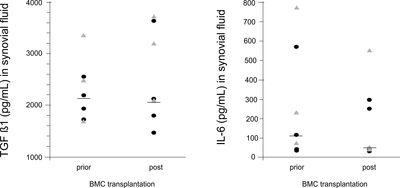
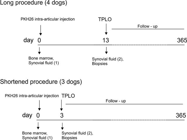
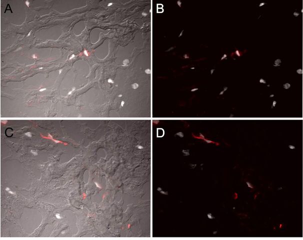
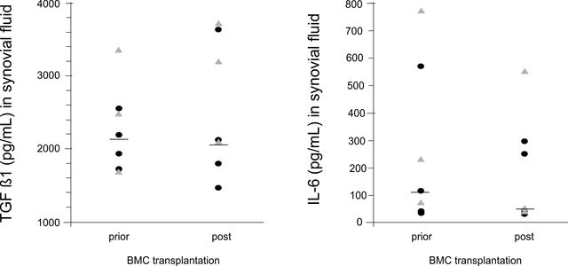
Current research indicates that exogenous stem cells may accelerate reparative processes in joint disease but, no previous studies have evaluated whether bone marrow cells (BMCs) target the injured cranial cruciate ligament (CCL) in dogs. The objective of this study was to investigate engraftment of BMCs following intra-articular injection in dogs with spontaneous CCL injury. Autologous PKH26-labelled BMCs were injected into the stifle joint of eight client-owned dogs with CCL rupture. The effects of PKH26 staining on cell viability and PKH26 fluorescence intensity were analysed in vitro using a MTT assay and flow cytometry. Labelled BMCs in injured CCL tissue were identified using fluorescence microscopy of biopsies harvested 3 and 13 days after intra-articular BMC injection. The intensity of PKH26 fluorescence declines with cell division but was still detectable after 16 days. Labelling with PKH26 had no detectable effect on cell viability or proliferation. Only rare PKH26-positive cells were present in biopsies of the injured CCL in 3/7 dogs and in synovial fluid in 1/7 dogs. No differences in transforming growth factor-beta1, and interleukin-6 before and after BMC treatment were found and no clinical complications were noted during a 1 year follow-up period. In conclusion, BMCs were shown to engraft to the injured CCL in dogs when injected into the articular cavity. Intra-articular application of PKH26-labelled cultured mesenchymal stem cells is likely to result in higher numbers of engrafted cells that can be tracked using this method in a clinical setting.
Item Type: |
Journal Article (Original Article) |
|---|---|
Division/Institute: |
05 Veterinary Medicine > Department of Clinical Veterinary Medicine (DKV) 05 Veterinary Medicine > Department of Clinical Veterinary Medicine (DKV) > Small Animal Clinic > Small Animal Clinic, Surgery 05 Veterinary Medicine > Department of Clinical Veterinary Medicine (DKV) > Small Animal Clinic |
UniBE Contributor: |
Linon, Elisa, Spreng, David Emmanuel, Rytz, Ulrich, Forterre, Simone |
Subjects: |
500 Science 600 Technology > 610 Medicine & health |
ISSN: |
1090-0233 |
Publisher: |
Elsevier |
Language: |
English |
Submitter: |
Simone Forterre |
Date Deposited: |
02 Feb 2015 15:21 |
Last Modified: |
02 Mar 2023 23:25 |
Publisher DOI: |
10.1016/j.tvjl.2014.08.031 |
PubMed ID: |
25261229 |
BORIS DOI: |
10.7892/boris.61314 |
URI: |
https://boris.unibe.ch/id/eprint/61314 |
Actions (login required)
 |
Edit item |