
Haefeli, Pascal Cyrill; Steppacher, Simon Damian; Babst, Doris; Siebenrock, Klaus Arno; Tannast, Moritz (2015). An Increased Iliocapsularis-to-rectus-femoris Ratio Is Suggestive for Instability in Borderline Hips. Clinical orthopaedics and related research, 473(12), pp. 3725-3734. Springer 10.1007/s11999-015-4382-y
![[img]](https://boris.unibe.ch/style/images/fileicons/text.png) |
Text
art%3A10.1007%2Fs11999-015-4382-y.pdf - Published Version Restricted to registered users only Available under License Publisher holds Copyright. Download (1MB) |
|
|
Text
Iliocapsularis_BORIS_CopyeditedVersion_2016-03-22.pdf - Accepted Version Available under License Publisher holds Copyright. Download (225kB) | Preview |
|
|
Text (Table 1)
Table1_Demorgraphics.pdf - Accepted Version Available under License Publisher holds Copyright. Download (58kB) | Preview |
|
|
Text (Table 2)
Table2_Measurements.pdf - Accepted Version Available under License Publisher holds Copyright. Download (56kB) | Preview |
|
|
Text (Table 3)
Table3_2x2Tables_SensSpezPPVNPVAcc.pdf - Accepted Version Available under License Publisher holds Copyright. Download (66kB) | Preview |
|
![[img]](https://boris.unibe.ch/75703/12/Figure1_AngleMeasurements.jpg)
|
Image (Figure 1)
Figure1_AngleMeasurements.jpg - Accepted Version Available under License Publisher holds Copyright. Download (1MB) | Preview |
|
![[img]](https://boris.unibe.ch/75703/13/Figure2_IliocapsularisAnatomy%20Kopie.jpg)
|
Image (Figure 2)
Figure2_IliocapsularisAnatomy Kopie.jpg - Accepted Version Available under License Publisher holds Copyright. Download (1MB) | Preview |
|
![[img]](https://boris.unibe.ch/75703/14/Figure3_Measurements%20Kopie.jpg)
|
Image (Figure 3)
Figure3_Measurements Kopie.jpg - Accepted Version Available under License Publisher holds Copyright. Download (1MB) | Preview |
|
![[img]](https://boris.unibe.ch/75703/15/Figure4_CSA_3GroupsBoxplots%20Kopie.jpg)
|
Image (Figure 4)
Figure4_CSA_3GroupsBoxplots Kopie.jpg - Accepted Version Available under License Publisher holds Copyright. Download (1MB) | Preview |
|
![[img]](https://boris.unibe.ch/75703/16/Figure5_2x3_BorderlineCases%20Kopie.jpg)
|
Image (Figure 5)
Figure5_2x3_BorderlineCases Kopie.jpg - Accepted Version Available under License Publisher holds Copyright. Download (1MB) | Preview |

BACKGROUND
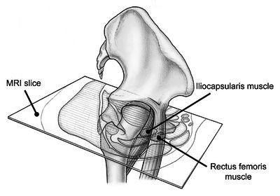
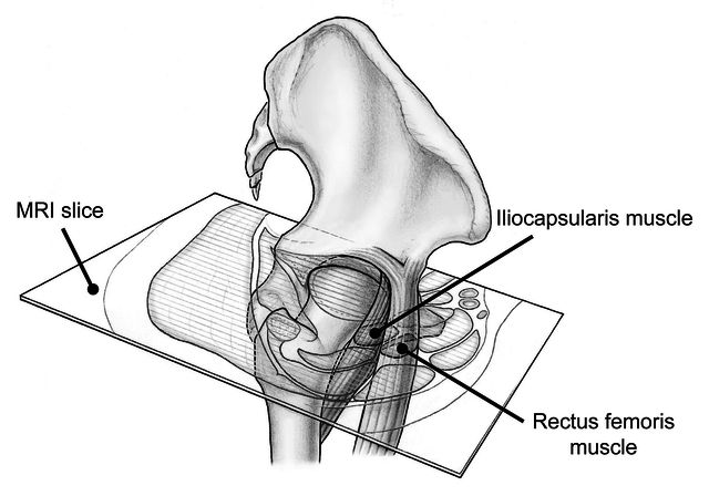
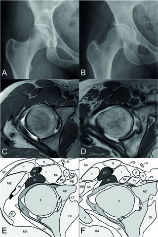
The iliocapsularis muscle is an anterior hip structure that appears to function as a stabilizer in normal hips. Previous studies have shown that the iliocapsularis is hypertrophied in developmental dysplasia of the hip (DDH). An easy MR-based measurement of the ratio of the size of the iliocapsularis to that of adjacent anatomical structures such as the rectus femoris muscle might be helpful in everyday clinical use.
QUESTIONS/PURPOSES
We asked (1) whether the iliocapsularis-to-rectus-femoris ratio for cross-sectional area, thickness, width, and circumference is increased in DDH when compared with hips with acetabular overcoverage or normal hips; and (2) what is the diagnostic performance of these ratios to distinguish dysplastic from pincer hips?
METHODS
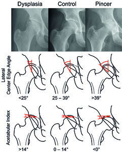
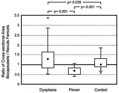
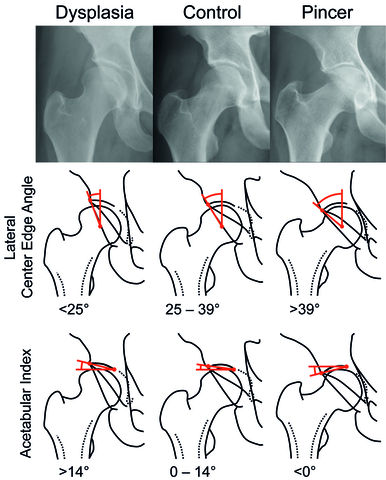
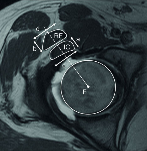
We retrospectively compared the anatomy of the iliocapsularis muscle between two study groups with symptomatic hips with different acetabular coverage and a control group with asymptomatic hips. The study groups were selected from a series of patients seen at the outpatient clinic for DDH or femoroacetabular impingement. The allocation to a study group was based on conventional radiographs: the dysplasia group was defined by a lateral center-edge (LCE) angle of < 25° with a minimal acetabular index of 14° and consisted of 45 patients (45 hips); the pincer group was defined by an LCE angle exceeding 39° and consisted of 37 patients (40 hips). The control group consisted of 30 asymptomatic hips (26 patients) with MRIs performed for nonorthopaedic reasons. The anatomy of the iliocapsularis and rectus femoris muscle was evaluated using MR arthrography of the hip and the following parameters: cross-sectional area, thickness, width, and circumference. The iliocapsularis-to-rectus-femoris ratio of these four anatomical parameters was then compared between the two study groups and the control group. The diagnostic performance of these ratios to distinguish dysplasia from protrusio was evaluated by calculating receiver operating characteristic (ROC) curves and the positive predictive value (PPV) for a ratio > 1. Presence and absence of DDH (ground truth) were determined on plain radiographs using the previously mentioned radiographic parameters. Evaluation of radiographs and MRIs was performed in a blinded fashion. The PPV was chosen because it indicates how likely a hip is dysplastic if the iliocapsularis-to-rectus-femoris ratio was > 1.
RESULTS
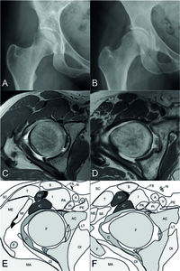
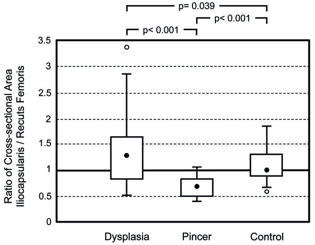
The iliocapsularis-to-rectus-femoris ratio for cross-sectional area, thickness, width, and circumference was increased in hips with radiographic evidence of DDH (ratios ranging from 1.31 to 1.35) compared with pincer (ratios ranging from 0.71 to 0.90; p < 0.001) and compared with the control group, the ratio of cross-sectional area, thickness, width, and circumference was increased (ratios ranging from 1.10 to 1.15; p ranging from 0.002 to 0.039). The area under the ROC curve ranged from 0.781 to 0.852. For a one-to-one iliocapsularis-to-rectus-femoris ratio, the PPV was 89% (95% confidence interval [CI], 73%-96%) for cross-sectional area, 77% (95% CI, 61%-88%) for thickness, 83% (95% CI, 67%-92%) for width, and 82% (95% CI, 67%-91%) for circumference.
CONCLUSIONS
The iliocapsularis-to-rectus-femoris ratio seems to be a valuable secondary sign of DDH. This parameter can be used as an adjunct for clinical decision-making in hips with borderline hip dysplasia and a concomitant cam-type deformity to identify the predominant pathology. Future studies will need to prove this finding can help clinicians determine whether the borderline dysplasia accounts for the hip symptoms with which the patient presents.
LEVEL OF EVIDENCE
Level III, prognostic study.
Item Type: |
Journal Article (Original Article) |
|---|---|
Division/Institute: |
04 Faculty of Medicine > Department of Orthopaedic, Plastic and Hand Surgery (DOPH) > Clinic of Orthopaedic Surgery |
UniBE Contributor: |
Haefeli, Pascal, Steppacher, Simon Damian, Siebenrock, Klaus-Arno, Tannast, Moritz |
Subjects: |
600 Technology > 610 Medicine & health |
ISSN: |
0009-921X |
Publisher: |
Springer |
Language: |
English |
Submitter: |
Fabian Röthlisberger |
Date Deposited: |
22 Mar 2016 11:44 |
Last Modified: |
05 Dec 2022 14:51 |
Publisher DOI: |
10.1007/s11999-015-4382-y |
PubMed ID: |
26088766 |
BORIS DOI: |
10.7892/boris.75703 |
URI: |
https://boris.unibe.ch/id/eprint/75703 |
Actions (login required)
 |
Edit item |